Emergency: for a life-threatening emergency, call 911 or go to the nearest emergency department.

Monday–Friday, 8:30 AM–5:00 PMSe habla españolCall 404-446-4424

Care & Conditions

Conditions and services designed around your next step

Our doctors and rehabilitation team evaluate concerns of the brain, spine, and peripheral nerves, then explain the diagnostic, non-surgical, interventional, or surgical options that may be appropriate.

Georgia Brain and Spine Center Johns Creek office exterior
General information only: The material below is educational and does not replace a personal evaluation or medical advice. Sudden or severe symptoms need prompt medical attention.

Clear answers for complex brain and balance concerns.

Symptoms involving the brain, dizziness, or balance can feel unsettling. Our surgeons explain what an evaluation may involve and help you understand the appropriate next step.

Dr. Elias Dagnew smiling in a navy suit and gold tie
Brain tumors

Brain tumors are abnormal growths in or around the brain that may be benign or malignant. Persistent headaches, seizures, unexplained nausea, vision or hearing changes, memory changes, balance problems, or personality changes deserve medical attention.

Care discussions may include imaging, observation for some tumors, biopsy, or surgery when appropriate.

Brain bleeds

Subdural, epidural, and intracerebral hemorrhages are types of bleeding in or around the brain. Sudden severe headache, sudden weakness or numbness, slurred speech, confusion, or loss of consciousness are emergency warning signs.

Hydrocephalus and normal pressure hydrocephalus (NPH)

Hydrocephalus is a buildup of cerebrospinal fluid (CSF) in the brain that enlarges the ventricles where CSF is stored. It can occur because an obstruction blocks the normal flow of CSF — obstructive hydrocephalus — or because the brain cannot reabsorb CSF as quickly as it is produced — communicating hydrocephalus. Symptoms may include headaches, nausea and vomiting, double vision, lethargy, and seizures. Hydrocephalus often presents at a young age and may be associated with some birth defects.

Normal pressure hydrocephalus (NPH) is a communicating form that usually appears in adults over 60. Its classic triad is memory problems, difficulty walking, and urinary incontinence. Identifying NPH matters because it is a treatable condition and one of the few reversible causes of dementia-like symptoms.

Evaluation includes a comprehensive neurological examination and appropriate imaging. In some cases a lumbar puncture, or a lumbar drain with a formal physical-therapy gait assessment, helps confirm the diagnosis and show whether surgery may help. Because other conditions can mimic hydrocephalus, a thorough work-up matters. Treatment may include a shunt that drains CSF into the abdominal cavity for the body to reabsorb, or an endoscopic third ventriculostomy for some obstructive cases. Shunts placed years earlier can develop a blockage or infection and may need revision.

Read the full hydrocephalus and NPH guide

Chiari malformation

A Chiari malformation occurs when the lower part of the skull cavity, called the posterior fossa, is crowded. The crowding pushes the cerebellar tonsils down through the opening at the bottom of the skull into the spinal canal. This can press on the brain stem and spinal cord and block the flow of cerebrospinal fluid from the brain to the spine.

Headaches made worse by coughing, laughing, crying, straining, or bending are common. Other symptoms can include weakness or numbness in the arms or legs, balance or gait problems, difficulty swallowing or hoarseness, ringing in the ears, dizziness, blurred vision, problems with concentration, and snoring. Many Chiari malformations cause no symptoms and are found incidentally on imaging studies.

Evaluation begins with a neurological examination and appropriate imaging, which may include MRI of the brain and spine and, in some cases, a CINE MRI to assess cerebrospinal fluid flow at the area of compression. Mild or absent symptoms may be followed with neurological check-ups and serial MRI. When symptoms are significant, a posterior fossa decompression can relieve pressure by removing a portion of the back of the skull and, at times, the back of the upper neck vertebrae.

Read the full Chiari malformation guide

Traumatic brain injury, contusions, and concussions

Traumatic brain injury is a broad diagnosis that ranges from concussion — also called mild traumatic brain injury — to severe injury. A concussion is a temporary neurological change such as loss of consciousness, confusion, distractibility, or memory problems after a head injury, without clear structural damage to the brain. Bruising of the brain is called a contusion. Severe traumatic brain injury involves more significant damage and requires care in an intensive care unit.

After a head injury, some people develop post-concussive symptoms: dizziness, headaches, blurred vision, balance problems, ringing in the ears, memory and concentration difficulty, and impaired judgment.

Early diagnosis and management matter. Most concussions and contusions are treated with supportive measures, while severe injuries may require surgery. Our concussion program pairs neurosurgeon-led evaluation with cognitive, vestibular, and balance assessment and individualized rehabilitation.

Vertigo & balance disorders

Spinning sensations, lightheadedness, unsteadiness, frequent stumbles, or nausea can affect daily life. Our team can discuss neurological evaluation and rehabilitation options for dizziness, balance, gait, and fall prevention.

Emergency: for sudden severe headache, weakness, trouble speaking, loss of consciousness, or loss of bladder or bowel control, call 911 or go to the nearest emergency department.

Neck and arm symptoms deserve a careful look.

Cervical conditions can contribute to neck pain, radiating arm symptoms, hand clumsiness, or gait changes. Our surgeons will explain which diagnostic and treatment conversations may be useful.

Meet our interventional spine specialist
Dr. Michael Hartman smiling in a white coat over a blue shirt and patterned tie
Cervical disc herniation

A disc in the neck can bulge or rupture and irritate nearby nerves. Symptoms may include neck pain into the shoulder, arm, or hand, with numbness, tingling, or weakness.

Cervical stenosis

Narrowing of the spinal canal in the neck can put pressure on the spinal cord or nerves. Stiffness, localized pain, and radiating numbness or weakness may be present.

Cervical myelopathy

Spinal cord compression in the neck needs prompt specialist evaluation. Difficulty with fine-motor tasks, dropping items, gait instability, or broad weakness are important symptoms to discuss.

Targeted conversations for mid- and low-back symptoms.

Back and leg symptoms can have several causes. A careful evaluation can help distinguish when physical therapy, imaging, injections, decompression, or a surgical opinion may be appropriate.

Woman in black athletic clothing performing a lunge beside a pond in an autumn park
Thoracic and lumbar stenosis

Thoracic stenosis can cause mid-back pain, band-like chest or abdominal discomfort, numbness, or leg weakness. Lumbar stenosis may cause lower-back and leg pain, cramping, or heaviness that worsens with walking or standing and improves with sitting or leaning forward.

Low back pain and sciatica

Low back pain and sciatica are among the most common reasons people seek medical care. About 90% of people improve within a month with little or no treatment. For others, back pain becomes severe, disabling, and persistent without intervention. Sciatica — pain shooting down the leg — is a sign of spinal nerve root compression.

These symptoms are nonspecific and the cause can be difficult to identify. Possible sources include muscle strain, degenerative disc disease, disc herniation, osteoarthritis, bone spurs, narrowing of the lumbar spinal canal, spondylolisthesis, inflammation of the sacroiliac joint, or infection of the bone. Imaging such as X-ray, CT, or MRI helps confirm the diagnosis and rule out red-flag conditions including infection, cancer, spinal fracture, and critical narrowing of the spinal canal.

We try conservative care first: physical therapy, analgesics, muscle relaxants, and spinal injections such as epidural, trigger-point, and lumbar facet injections, which our surgeons can perform in our office. When non-operative options are exhausted, surgery is tailored to your symptoms and the anatomy of your spine — lumbar laminectomy, removal of a disc herniation, fusion with pedicle screws, or a combination — using minimally invasive or traditional open techniques as appropriate.

Degenerative disc disease, osteoarthritis, and spondylosis

These broad terms describe age-related degeneration of the spinal column. Discs can dry out and tear, spinal joints can become overgrown and lax, and bone spurs and thickened ligament can develop. These changes can make the back unstable and cause pain, or create pressure on nerve roots and cause pain that radiates down the legs.

There is no one-size-fits-all solution, and some degree of change is a normal part of aging. We use your medical history, physical examination, and imaging to understand the type of pain you are experiencing and to locate the spinal abnormality responsible. Conservative non-surgical care is tried first. When surgery is needed, decompression addresses nerve-root pressure from thickened ligament and bone, while fusion addresses instability; both are frequently performed together.

Spondylolisthesis

Spondylolisthesis is a vertebra slipping forward over the one below it. It can cause back pain, stiffness, and difficulty standing or walking, and may narrow the spinal canal or compress nerve roots.

Evaluation includes examination and imaging to assess the degree of slip, stability, and any pressure on the nerves. Care may begin with physical therapy and activity guidance, with surgery considered when symptoms persist or the spine is unstable.

Tumors, fractures, and pain after spine surgery.

Some spinal conditions need a more specialized evaluation — tumors involving the spinal cord or spinal column, fractures that may destabilize the spine, and persistent pain after previous spine surgery. Our surgeons explain the imaging, the options, and what a tailored plan may involve.

Meet our surgeon with fellowship training in complex spinal surgical oncology
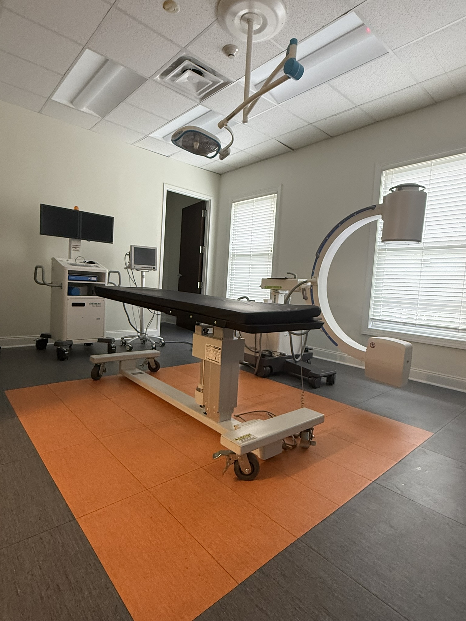
Georgia Brain and Spine procedure room with a patient table and C-arm imaging equipment
Spinal fractures

Spinal fractures are bone injuries that can occur in the cervical, thoracic, or lumbar spine, often after significant trauma such as a car accident, a fall down stairs, or a fall from standing. Conditions such as cancer and osteoporosis can weaken bone and predispose you to fracture after a minor injury. Fractures are classified by which bone is injured, the orientation of the fracture line, whether the spinal column is unstable, and whether the spinal cord or nerve roots are involved.

Minor fractures in a stable spinal column with no injury to the spinal cord or nerve roots can often be managed with an external brace. Unstable fractures, or those with damage to the spinal cord, require urgent surgery. Osteoporotic compression fractures may be treated with vertebroplasty or kyphoplasty, minimally invasive outpatient procedures that inject cement into the bone to restore strength and stability and can greatly reduce pain.

Read the full spinal fracture guide

Failed back syndrome

Some people continue to have chronic pain after spine surgery. This is called failed back syndrome. Every case is unique; possible causes include scar tissue, recurrent or persistent disc disease, continued narrowing of the spinal canal, mechanical instability, non-fusion of the spine, nerve damage, infection, or improper alignment of the spinal column.

Finding the precise source of pain is the key. Evaluation includes a thorough history, examination, and imaging. Non-surgical treatment — physical therapy, analgesics, and spinal injections — is tried first. If those measures do not help or are not an option, surgery may be designed to decompress narrow areas around the nerve roots, stabilize and fuse unstable bone, and in some cases use chronic pain approaches such as neuromodulation or spinal cord stimulation.

Tumors of the spinal cord and spinal column

Tumors can arise within or spread to the spinal cord or spinal column. As they grow they can destroy structurally important parts of the spine, leading to pain and mechanical instability, or compress and invade the spinal cord and nerve roots, causing difficulty walking, weakness, numbness, or loss of bladder or bowel control. Tumors of the spine are generally classified by location and aggressiveness.

Diagnosis is based on history, physical examination, and imaging such as CT and MRI. Treatment depends on the type of tumor, its location, and whether it is causing symptoms. Some benign tumors can be monitored and may never grow or cause symptoms. Aggressive tumors often benefit from surgery to remove them completely, and extensive resections may require complex reconstruction of the spinal column.

Nerve symptoms in the hands and arms.

Numbness, tingling, nighttime pain, and hand weakness can arise when a peripheral nerve is compressed. Our team can evaluate the nerve, explain what testing may be useful, and discuss treatment options.

Man in athletic clothing stretching one arm across his chest outdoors
Carpal tunnel syndrome

Carpal tunnel syndrome means added pressure on the median nerve around the wrist. The nerve passes through a tunnel inside the bones of the hand with a ligament across the top. Repetitive use of the hand can cause inflammation and thickening of that ligament, narrowing the tunnel and pressing on the nerve. This can cause hand pain, tingling, numbness, and, if untreated, weakness of the hand muscles.

Treatment starts with avoiding the repetitive activity that may be contributing, and a wrist brace to keep the wrist in a neutral position. A short course of anti-inflammatory medication or a cortisone injection into the carpal tunnel helps some people. When non-surgical options do not provide relief, surgery opens and releases the tunnel by cutting the ligament to take pressure off the nerve. The minimally invasive approach allows for a rapid recovery and is usually very successful in improving symptoms when performed by an experienced neurosurgeon.

Ulnar nerve entrapment

The ulnar nerve travels down the inner part of the arm, crossing the elbow and the wrist to supply the inner part of the hand. Entrapment can compress the nerve near the elbow — the cubital tunnel — or at the wrist, causing pain, tingling, numbness, and sometimes weakness in the hand. Diagnosis includes a detailed neurological examination with EMG and nerve conduction studies.

We begin by limiting the repetitive activity that may be contributing, including direct pressure on the elbow and wrist, and may brace the elbow or wrist in a neutral position. A short course of anti-inflammatory medication and physical therapy is often recommended. If non-surgical treatment does not achieve the result you need, surgery can open and release the nerve where it is entrapped; this outpatient procedure uses a small incision to free the nerve from the tissues compressing it.

Emergency: for sudden severe headache, weakness, trouble speaking, loss of consciousness, or loss of bladder or bowel control, call 911 or go to the nearest emergency department.

More ways to evaluate and treat spine, balance, and pain concerns.

Our doctors and rehabilitation team bring advanced imaging, physical therapy, balance care, interventional procedures, and surgical consultation together to help plan a next step.

Weight-bearing MRI

Our Esaote Brio weight-bearing MRI can image the spine while a person is standing or sitting. Unlike traditional flat-bed MRI, this approach evaluates the spine under natural load and may help the care team assess findings that are less evident while lying down.

The study is one part of a complete clinical evaluation. Your physician will explain whether this imaging approach may be useful for your symptoms and treatment planning.

  • Standing or seated imaging options for selected spine evaluations
  • Specialist review in the context of your symptoms and examination
  • Available at the Johns Creek office

Physical Therapy & Rehabilitation

Our Doctors of Physical Therapy create individualized programs for spine conditions, chronic pain, orthopedic concerns, and pre- or post-surgical rehabilitation. Care may focus on spinal stabilization, movement, strength, education, and a practical plan for daily activity.

Recommendations are based on an individual evaluation, goals, and current condition rather than a one-size-fits-all program.

  • Spinal stabilization and rehabilitation planning
  • Pre- and post-surgical physical therapy conversations
  • Care coordinated with the Georgia Brain & Spine clinical team

Concussion Care

A concussion or mild traumatic brain injury can follow a bump, blow, or jolt to the head. Headache, dizziness, difficulty with balance, memory, focus, or concentration, brain fog, fatigue, sleep changes, light or sound sensitivity, and mood changes are reasons to consider an evaluation.

Our comprehensive concussion program in Metro Atlanta is led by Elias Dagnew, MD, FACS, a Board-Certified Neurosurgeon with extensive experience in the diagnosis and management of traumatic brain injury. The program brings specialist evaluation and individualized therapy together, rather than ending with a diagnosis.

Care includes detailed assessment of cognitive function, including memory, focus, and concentration; the vestibular system, which supports orientation and movement; and balance. Working with our rehabilitation team, patients receive a tailored concussion-therapy plan aimed at helping them return to daily life, work, and activity at an appropriate pace.

Concussion care is generally non-surgical. Dr. Dagnew also has experience in non-surgical and surgical management of other traumatic brain injuries when indicated. An individual evaluation guides the appropriate care pathway; goals and recovery timelines vary.

  • Neurosurgeon-led evaluation of concussion and traumatic brain injury concerns
  • Detailed cognitive, vestibular, and balance assessment
  • Individualized concussion therapy and rehabilitation with practical return-to-activity guidance

Vertigo, Balance & Fall Prevention

Dizziness, vertigo, gait changes, and frequent stumbles can affect confidence and daily routines. Our rehabilitation team offers vestibular-retraining, balance, gait, and fall-prevention conversations alongside neurological evaluation when appropriate.

The team can discuss symptoms such as spinning sensations, lightheadedness, unsteadiness, or nausea and help identify a next step for evaluation and rehabilitation.

  • Vestibular-rehabilitation and balance-care conversations
  • Gait and fall-prevention support, including AgeSteady resources
  • Coordination with concussion and neurological care when appropriate

DRX9000 Spinal Decompression

The DRX9000 is an advanced, computer-controlled traction system designed to gently relieve pressure on spinal discs. It is a non-invasive option that may be considered for selected disc-related spine concerns after evaluation.

Our team can discuss whether spinal decompression, physical therapy, injections, or another pathway may be appropriate for your situation.

  • Computer-controlled traction under clinical guidance
  • A non-invasive option for selected patients
  • Part of a broader non-surgical care discussion

Interventional Pain Management

Interventional procedures can help physicians clarify the source of pain or deliver targeted treatment when appropriate. Dr. Hartman may discuss image-guided options as part of a broader diagnostic and care plan.

Whether a procedure is appropriate depends on your diagnosis, symptoms, imaging, and clinical evaluation.

  • Epidural steroid injections for selected neck, back, or radiating pain concerns
  • Facet-joint and medial-branch blocks to assess or address selected spinal-joint pain patterns
  • Platelet-rich plasma (PRP) injections may be discussed for selected tendon, ligament, and spinal-structure concerns

Surgery & Procedures

Our board-certified neurosurgeons consider surgery when it may offer an appropriate next step after careful evaluation. They discuss the procedure, alternatives, and questions that matter before a decision is made.

Potential conversations can include cervical, thoracic, and lumbar procedures; brain surgery; and peripheral nerve procedures when indicated.

  • ACDF, cervical arthroplasty, laminectomy, microdiscectomy, fusion, and kyphoplasty may be discussed for selected spine conditions
  • Brain-tumor, hydrocephalus, and brain-bleed treatment planning when indicated
  • Carpal and cubital tunnel release / ulnar nerve neurolysis conversations for selected peripheral nerve conditions

Understand your surgical options

Explore Individual Procedures Choose one procedure for a focused explanation.

Understand the procedure. Discuss your options.

Our surgeons explain when a procedure may be appropriate, how it is generally performed, and what alternatives to consider. The descriptions below are educational; technique, suitability, risks, and recovery vary by person.

Learn about surgical approaches for conditions affecting the brain, spine, and peripheral nerves. This information is general education, not a personal treatment recommendation. Whether a procedure is appropriate, the technique used, and the outcome depend on each patient's condition and treatment plan.

Choose a surgical procedure

Explore 13 procedures by area of care. Choose one treatment to read its description.

Hydrocephalus & surgical treatment

Brain-bleed treatment planning

Emergency warning: If you suspect a brain bleed or are experiencing a medical emergency, call 911. Do not wait for an appointment or a response from the practice.

Cervical spine procedures

Lumbar & spinal decompression procedures

Cranial procedures & tumor care

Peripheral nerve procedures

Hydrocephalus & surgical treatment

Ventriculoperitoneal (VP) shunt placement

What it is: A VP shunt is a thin, flexible tube system, called a catheter, with a small control valve. It is surgically placed under the skin to divert excess cerebrospinal fluid (CSF) from the brain's fluid-filled cavities, or ventricles.

How it works: The catheter directs fluid into the abdominal cavity, or peritoneum, where the body can absorb it. The valve helps regulate drainage in response to pressure. This is intended to reduce excess fluid and pressure inside the skull and may help symptoms such as headaches, nausea, confusion, or balance difficulties. Shunt selection and placement vary according to the patient's needs; symptom improvement and pressure control are not guaranteed.

Request an appointment to discuss hydrocephalus treatment.

Brain-bleed treatment planning

An intracranial hemorrhage occurs when a blood vessel inside or around the brain ruptures, allowing blood to collect and press against brain tissue. Three types of brain bleeds described below differ in where blood collects:

  • Epidural hematoma: Blood collects between the skull and the tough outer brain lining, called the dura. It may expand rapidly after a head injury.
  • Subdural hematoma: Blood collects between the brain surface and its outer lining. An acute collection may develop quickly after severe trauma; a chronic collection may develop over weeks after a minor bump.
  • Intracerebral hemorrhage: Bleeding occurs within the brain tissue itself and may be associated with high blood pressure, vascular conditions, or trauma.

Emergency warning: If you suspect a brain bleed or are experiencing a medical emergency, call 911. Do not wait for an appointment or a response from the practice.

Treatment planning depends on the bleed's location, size, and severity. When surgical evacuation is needed to address pressure, the approaches below may be considered. The technique and extent of treatment vary by patient.

Burr-hole drainage

What it is: Burr-hole drainage uses small openings in the skull to access a blood collection. It may be considered for liquefied or chronic collections, such as a chronic subdural hematoma, when drainage is appropriate.

How it works: The surgeon creates one or more small, round openings through the skull so collected blood can be drained. Removing the collection is intended to reduce pressure on the brain. The number and location of openings and the drainage technique depend on the collection and the patient's treatment plan; this approach is not suitable for every bleed and does not guarantee relief or a particular recovery.

Request a non-emergency appointment to discuss brain-bleed care.

Craniotomy for hematoma evacuation

What it is: A craniotomy for hematoma evacuation involves temporarily removing a section of skull, called a bone flap, to access a blood collection. This approach may be used for larger, solid blood clots or acute bleeds when surgical removal is needed.

How it works: The opening gives the surgeon direct access to remove pooled blood and address active bleeding when possible. The procedure is intended to reduce pressure on the brain. The bone flap may be secured back in place, depending on the clinical situation. The extent of evacuation, bleeding control, and management of the bone flap are individualized rather than identical for every patient.

Request a non-emergency appointment to discuss hematoma treatment.

Cervical spine procedures

Anterior cervical discectomy and fusion (ACDF)

What it is: ACDF approaches the cervical spine through the front, or anterior, of the neck to remove a damaged or herniated disc pressing on the spinal cord or exiting nerve roots. It combines disc removal with fusion of the affected vertebrae.

How it works: The surgeon removes compressive disc material and may remove bone spurs. A spacer or graft may be placed in the disc space to support disc height, and a plate and screws may help hold the vertebrae steady while bone fusion develops over time. The procedure is intended to reduce nerve or spinal cord compression and may help arm pain, numbness, weakness, or balance problems related to cervical radiculopathy or myelopathy. The incision, graft, hardware, and technique vary; fusion and symptom improvement are not guaranteed.

Request an appointment to discuss ACDF and your cervical spine symptoms.

Cervical arthroplasty (artificial disc replacement)

What it is: Cervical arthroplasty is an artificial disc replacement procedure intended to preserve motion at a treated level in the neck. It may be an alternative to fusion for selected patients with cervical disc herniation or nerve compression.

How it works: Through an approach at the front of the neck, the surgeon removes the damaged disc and addresses nerve pressure. An artificial disc is placed in the disc space instead of intentionally fusing the adjacent bones. The implant is intended to support disc height and allow motion at the treated level. Device selection and technique depend on the patient's anatomy and condition; the procedure does not guarantee symptom relief, natural neck flexibility, or reduced stress on neighboring spinal levels.

Request an appointment to discuss whether cervical disc replacement may be appropriate.

Lumbar & spinal decompression procedures

Microdiscectomy

What it is: Microdiscectomy is a targeted procedure to remove herniated or bulging disc material in the lower back, or lumbar spine, that is pressing on a spinal nerve root.

How it works: Through an incision in the lower back, the surgeon accesses the affected disc and removes the portion compressing the nerve, with the aim of preserving as much unaffected disc and bone as possible. Magnification or endoscopic techniques may be used; the approach and handling of the nerve vary with the individual case. Decompression is intended to address radiating leg pain, or sciatica, numbness, or weakness. Symptom relief and the pace of recovery vary and are not guaranteed.

Request an appointment to discuss a lumbar disc problem.

Laminectomy (spinal decompression)

What it is: Laminectomy is a decompression procedure intended to reduce pressure on the spinal cord or spinal nerves caused by spinal stenosis, which is narrowing of the spinal canal, or by bone spurs, also called osteophytes.

How it works: Through an approach along the back of the spine, the surgeon removes part or all of the lamina, the bony arch forming the back of the spinal canal. Thickened ligaments or bone spurs may also be removed. This creates more space for compressed nerves and may help leg heaviness, cramping, pain, or numbness with standing or walking. The amount of bone and tissue removed is individualized, and improvement is not assured.

Request an appointment to discuss spinal stenosis and decompression.

Spinal fusion

What it is: Spinal fusion is intended to join and stabilize two or more vertebrae in the neck, mid-back, or lower back. It may be considered for instability associated with conditions such as spondylolisthesis, severe degenerative disc disease, or spinal trauma.

How it works: Bone graft material is placed between or along the affected vertebrae to encourage bone growth. Hardware such as rods, screws, or interbody cages may hold the bones in position while fusion develops. The goal is to support alignment and limit motion at the treated levels. The approach, graft, and hardware depend on the patient's condition; successful fusion, relief of pain, and correction of instability are not guaranteed.

Request an appointment to discuss spinal stability and fusion options.

Kyphoplasty

What it is: Kyphoplasty is an image-guided procedure used for selected painful vertebral compression fractures, in which a spinal bone has collapsed. These fractures are commonly associated with osteoporosis or structural bone weakness.

How it works: Using X-ray guidance, the surgeon passes an instrument into the fractured vertebra. A balloon may be inflated to create a cavity and attempt to restore some of the lost vertebral height, then deflated and removed. Bone cement is placed in the cavity and hardens to help stabilize the fracture internally. The technique varies by case. Height restoration, pain relief, and rapid recovery are not guaranteed.

Request an appointment to discuss a vertebral compression fracture.

Cranial procedures & tumor care

Craniotomy for tumor resection

What it is: A craniotomy for tumor resection is an operation to access and remove or reduce a brain tumor, whether benign or malignant. A section of skull, called a bone flap, is removed to provide access to the treatment area.

How it works: The surgeon works through the opening to remove tumor tissue, balancing the extent of removal against the need to protect surrounding brain tissue and function. Microsurgical instruments and imaging or navigation guidance may be used as appropriate to the surgical plan. The bone flap may be reattached with fixation hardware after the tumor work. The approach, available guidance, extent of resection, and closure vary; complete removal and preservation of function cannot be guaranteed.

Request an appointment to discuss brain tumor surgical care.

Stereotactic brain biopsy

What it is: Stereotactic brain biopsy is a diagnostic procedure to collect a small tissue sample from a brain lesion or tumor. It may be considered when the target is deep within the brain or near sensitive areas.

How it works: MRI or CT images are used to plan a path to the target, and stereotactic guidance helps direct a narrow biopsy instrument through a small skull opening to obtain tissue. The sample is sent for pathology examination to help establish a diagnosis and guide treatment planning. The imaging, guidance method, and sampling technique vary by case. The procedure may obtain tissue without a larger open resection, but a definitive diagnosis is not guaranteed.

Request an appointment to discuss evaluation of a brain lesion.

Peripheral nerve procedures

Carpal tunnel release

What it is: Carpal tunnel release is a procedure intended to reduce pressure on the median nerve as it passes through the wrist into the hand. It may be performed on an outpatient basis.

How it works: Compression beneath the transverse carpal ligament, which forms the roof of the tunnel, can cause pain, tingling, or numbness in the thumb, index, and middle fingers. The surgeon divides this ligament to increase space within the tunnel and reduce pressure on the nerve. The surgical approach varies according to the patient's needs. Sensation, strength, and symptoms may improve, but recovery of nerve function and prevention of lasting nerve damage are not guaranteed.

Request an appointment to discuss carpal tunnel symptoms.

Cubital tunnel release & ulnar nerve neurolysis (transposition when appropriate)

What it is: Cubital tunnel release and ulnar nerve neurolysis are procedures intended to relieve compression of the ulnar nerve at the elbow, near the area often called the funny bone. Compression may cause numbness, tingling, or weakness in the ring and little fingers and reduced grip strength.

How it works: Through an approach near the elbow, the surgeon releases tight tissue bands or ligaments around the nerve; this freeing of the nerve is called neurolysis. If the nerve is prone to slipping or irritation as the elbow bends, it may be moved to the front of the elbow, called ulnar nerve transposition. This is intended to reduce stretch or compression. Transposition is not necessary for every patient, and the technique and degree of symptom improvement vary.

Request an appointment to discuss ulnar nerve symptoms and treatment options.
Ready for a next step?
Speak with our team about appointments and practical next steps.